

Our Services
Human-Quality Diagnostic Imaging for Dogs & Cats
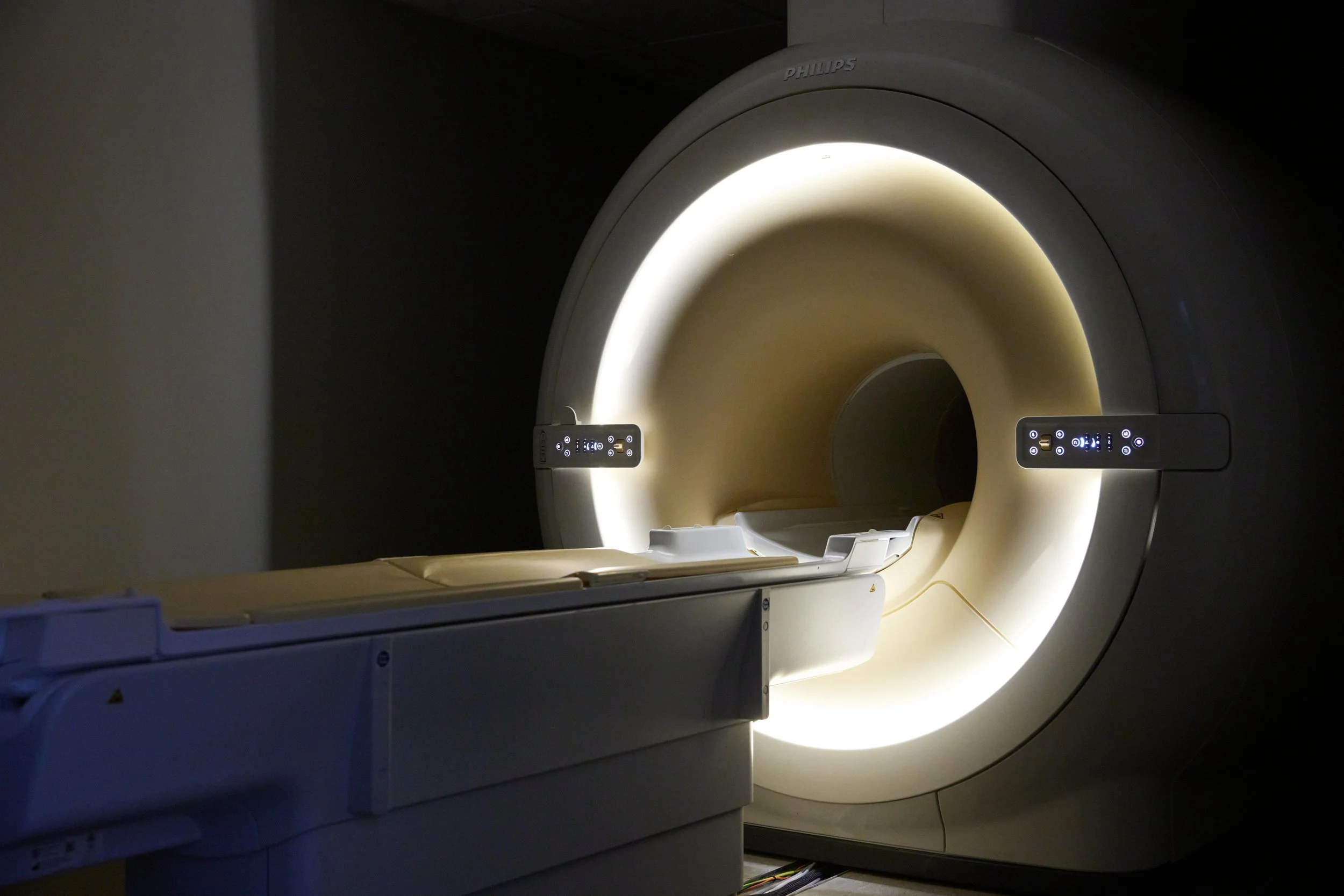
Board-certified veterinary radiologists using human-grade 3T MRI, 128-slice CT, ultrasound, and echocardiography — with same-day results and a direct call on every case.
Advanced Diagnostic Imaging
Human-grade equipment reveals what standard exams and X-rays cannot. Every study is read by a board-certified veterinary radiologist and includes a direct call to discuss findings.


Beyond Imaging
From neurological workups to rapid cytology, our specialized services complete the diagnostic picture — often in a single visit.


Radiotherapy & Specialized Treatment
Through our sister companies, we offer specialized treatment at our Round Rock facility.
Decision support before you refer
Not sure which study a case needs? These tools map clinical signs to the right modality and make referral effortless.
Frequently Asked Questions
What diagnostic imaging services does Sage Veterinary Imaging offer?
Who interprets the imaging studies at Sage?
How quickly are results available?
Does my pet need anesthesia for imaging?
Where is Sage Veterinary Imaging located?
Explore 170+ Conditions We Diagnose
Browse by body system, symptom, or modality to find detailed imaging guidance for your patient or pet.
Sage Veterinary Imaging — board-certified veterinary radiology in Round Rock & Spring, Texas and Sandy, Utah. Imaging protocols are confirmed by our radiologists based on the patient and clinical question.