
The Evolution of Animal Imaging (1980s–Today)
Short on time? Download the easy-to-share education tool.

Over the last 40 years, veterinary medicine has quietly transformed.
Not because of new pills or surgeries, but because we can now see what used to be hidden in pet health. From blurry ultrasound pictures in the 1980s to today’s crisp MRI scans, diagnostic imaging has changed how we find and treat disease in animals.
But this evolution is more than just the latest technology. It’s the adoption of forward-thinking business models–and above all: the belief that pets deserve the same high-quality care as people.
At Sage Veterinary Imaging (SVI), we use ultrasound, CT, and MRI to deliver fast, detailed answers that help you have peace of mind when it comes to your furry friends.
Here’s a look at how it all started, and where we’re headed as experts in veterinary imaging and stewards of your pet’s health.
1980s: The Age of Limited Access
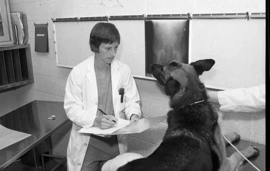
Dr. Gretchen Flo, a pioneer in veterinary orthopedics, early in her career reviewing X-rays with a patient. Photo courtesy of Michigan State University College of Veterinary Medicine
In the early 1980s, diagnostic imaging in veterinary medicine was still largely limited to radiographs (X-rays). Ultrasound was just beginning to emerge as a tool in academic settings, but its availability in general practice was rare.
Ultrasound units were big, expensive, and not very clear. Most general practices didn’t have access to them.
MRI and CT were only found in human hospitals. Vets had to work with teaching hospitals to use them, and even then, it wasn’t easy.
Diagnosing many internal problems felt like guesswork. Vets had to rely on symptoms and hunches. Sometimes, the only way to know what was wrong was to do surgery and look inside.
Veterinary radiology was still considered a niche specialty, and the idea of advanced outpatient imaging was years away.

Early radiograph from a 1988 case study of a Standard Poodle with chronic lameness. Images courtesy of Veterinary Radiology & Ultrasound / Wiley Online Library
1990s: The Era of Expansion
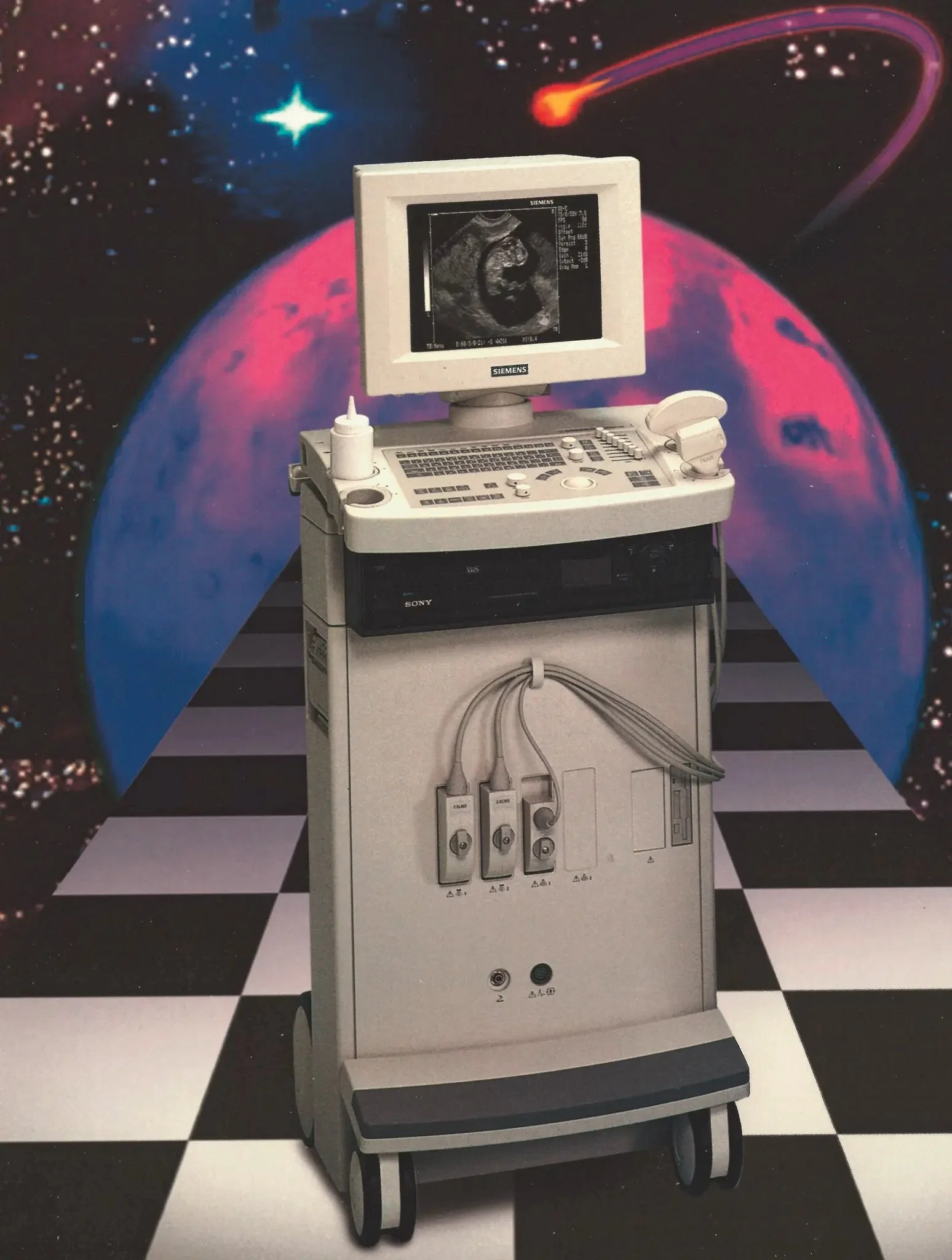
The SONOLINE Prima, launched in 1995, was Siemens’ first ultrasound system with digital workflow. Photo courtesy of Siemens Healthineers
As technology became more portable and cost-effective, things began to change in the 1990s. Imaging machines got smaller, cheaper, and easier to use.
Ultrasound began to enter general practice, especially in large practices. Mobile services popped up, with sonographers visiting different practices.
CT scanners trickled into major academic and referral centers. They were mostly used for bones and sinuses, and even then, only in special cases.
Veterinary MRIs remained limited to academia. Some animal hospitals tried using human machines after hours, but that brought challenges like safety and scheduling.
Meanwhile, pet owners were starting to ask more questions. They wanted answers. Vets felt the pressure to offer better diagnostics, just like doctors do for people.
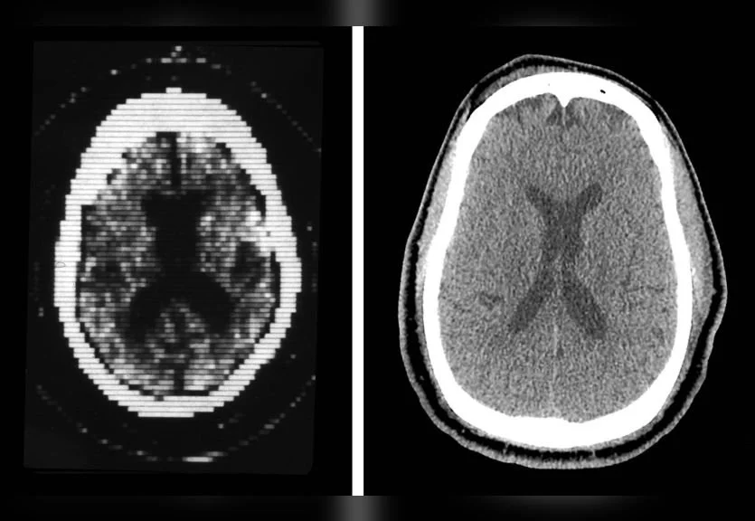
Side-by-side comparison of early CT imaging and modern scan quality. Image courtesy of Yale School of Medicine
2000s: The Rise of Advanced Diagnostic Imaging
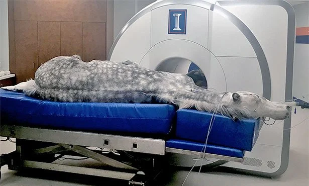
MRI scan of a horse at a veterinary teaching hospital. Photo courtesy of University of Illinois College of Veterinary Medicine
By the early 2000s, veterinary medicine was stepping into a new chapter. Specialty hospitals were growing. More vets were becoming board-certified. And imaging became a must-have.
Mobile imaging services grew rapidly. Experts traveled with machines, helping clinics without their own equipment. The images definitely helped, but sometimes lacked the detail needed to see more complex disease.
Veterinary neurologists and surgeons began to require imaging before treatment. CT and MRI went from “nice to have” to “need to have.”
Veterinary-specific MRI and CT units entered the market. These were typically low-field MRI systems (0.2T–0.5T) and 8- to 16-slice CT scanners; smaller and less powerful than human versions, but they got the job done.
Referral hospitals consolidated diagnostic and treatment services under one roof. But this new model created significant bottlenecks, as imaging demand far outpaced capacity.
As more pets began receiving MRIs, neurologic and orthopedic diagnostics reached new levels of accuracy. But clients often waited days or weeks for access—and prices continued to climb.
Mobile CT brings advanced imaging directly to patients, expanding access beyond specialty clinics. Photo courtesy of Mobile Pet Imaging
2010s: The Imaging Bottleneck
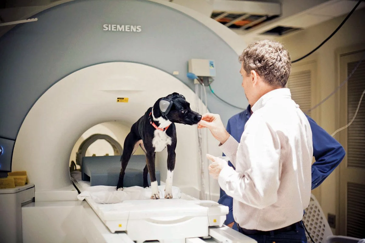
Emory researchers use fMRI to study canine cognition and behavior. Photo courtesy of Emory Magazine / Bryan Meltz
In the 2010s, imaging demand exploded. But access didn’t keep up.
More specialists were using MRI. More pet owners were asking for it. But there just weren’t enough machines (or trained radiologists) to go around.
Many general practices still didn’t have advanced imaging in-house. They had to refer patients, which meant more delays, more cost, and more stress for clients.
Specialty hospitals became overwhelmed. Long waitlists. Overworked teams. Slower care.
That’s when Sage Veterinary Imaging was born.
In 2016, we began by offering mobile ultrasound to clinics across Central Texas, bringing high-quality imaging straight to the patient. By 2020, we opened our first outpatient imaging center, built to deliver same-day MRI and CT scans without the delays and complexities of a referral hospital.
It was a new way forward: fast, clear answers that help veterinarians take action sooner.
2020s: The Rise of Outpatient Veterinary Imaging
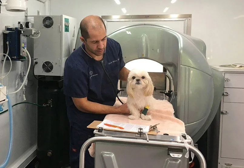
High-field MRI setup at Sage Veterinary Imaging. Photo by Kevin Vargo for Sage Veterinary Imaging
Today, outpatient imaging is rapidly reshaping the veterinary landscape, just as it did in human medicine two decades prior.
SVI and other centers now offer same-day MRI and CT with 3T and 128-slice technology.
Pet owners can schedule directly or work through their family vet. Either way, the process is smooth and simple.
Reports are usually delivered within 24 hours, often the same day. That means vets can make quicker decisions and get pets on the path to treatment sooner.
Outpatient centers take pressure off emergency and specialty teams. That gives them more time to focus on surgeries, critical cases, and the pets who need them most.
The result is a more balanced, efficient veterinary care ecosystem where diagnostics are timely, high-quality, and integrated into the broader workflow of general practice and referral care.
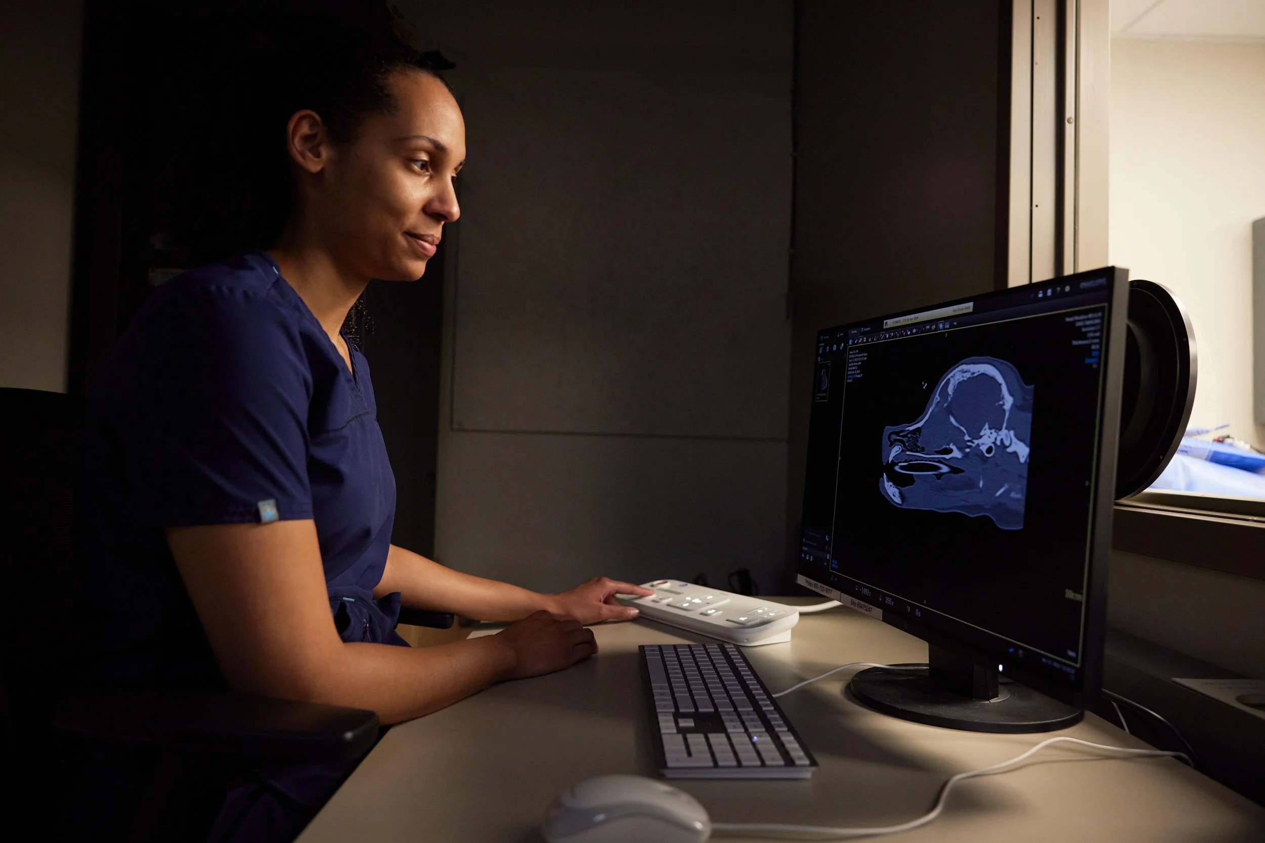
A board-certified veterinary radiologist reviews a CT scan as part of same-day diagnostic reporting. Photo by Kevin Vargo for Sage Veterinary Imaging
What's Next? Imaging in the 2030s
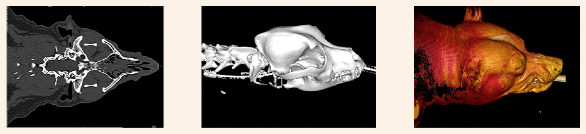
Imaging case study examples showcasing the advanced detail possible with modern CT and 3D reconstruction techniques. Images courtesy of Sage Veterinary Imaging

PET imaging supporting research for veterinary radiopharmaceuticals. Images courtesy of Antioxidants / MDPI
The next chapter in veterinary imaging is already taking shape. At SVI, we’re embracing new tools and technologies that make care even better—for pets, vets, and families.
AI-assisted scan reading is getting smarter. It helps radiologists work faster and with greater accuracy, so results come back quickly and with confidence.
3D and functional imaging are on the rise. These scans show what’s there and how it’s working, from blood flow to nerve function.
New tools will help pet owners see what we see. With clearer visuals and easier explanations, clients can better understand diagnoses and feel part of the care plan.
Diagnostic hubs will serve wider areas. Outpatient centers like ours will connect digitally with more clinics, helping pets get advanced care no matter where they live.
The future of imaging isn’t just faster and clearer; it’s smarter, more collaborative, and more integrated into everyday practice.
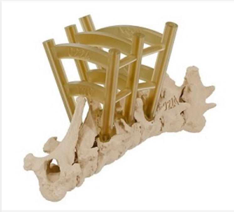
Image of 3D-printed drill guide for spine. Courtesy of The Royal School of Veterinary Studies, University of Edinburgh
Comprehensive Diagnostic Services at SVI
At Sage Veterinary Imaging, we’re more than an MRI and CT center. We provide a wide range of advanced imaging and diagnostic services—all under one roof—to support timely, accurate, and collaborative care.
Sage Veterinary Imaging’s services include:
3T MRI – High-resolution neuro, orthopedic, and soft tissue imaging with industry-leading detail
128-Slice CT – Rapid, full-body scanning ideal for trauma, bone, lung, and complex surgical planning
X-ray – Onsite digital radiography for rapid assessments and urgent case triage
Ultrasound & Echocardiography – Comprehensive abdominal and cardiac studies with same-day interpretation
Digital Cytology Lab – In-house rapid sample analysis to expedite diagnostics and reduce wait times
In-House Hematology Machines – Immediate bloodwork results to support imaging and anesthesia decisions
External Laboratory Testing – Access to specialized diagnostics including infectious disease panels and endocrine assays
Nuclear Scintigraphy for Feline Hyperthyroidism – Precise thyroid imaging using radiotracer techniques
Limited Nuclear Pharmaceutical Therapy – Targeted diagnostic and therapeutic applications using nuclear medicine
I-131 Therapy for Feline Hyperthyroidism – Definitive treatment using radioactive iodine administered on site
Synovetin OA for Canine Osteoarthritis – Intra-articular radiotherapeutic injection designed to relieve chronic joint pain
All of these services are designed to work together, making the diagnostic process smoother, faster, and more effective for vets and for your pets.
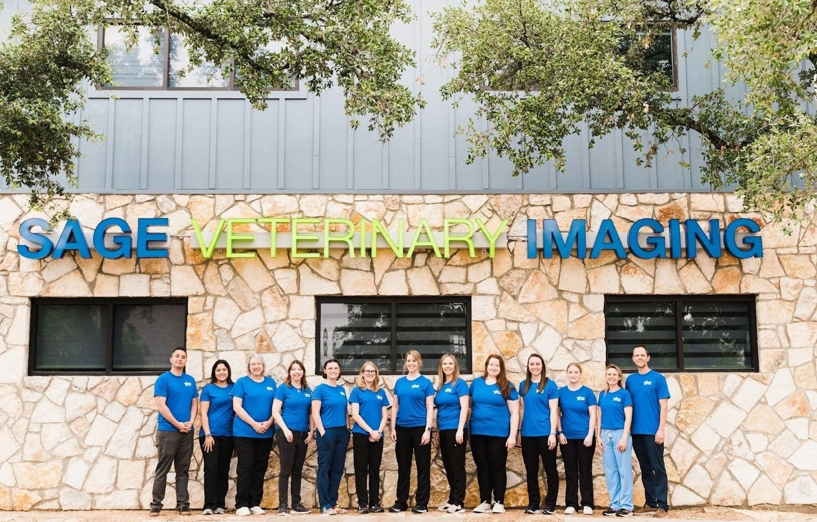
The Sage Veterinary Imaging team in Round Rock, Texas.
Final Thoughts
The story of animal imaging is one of progress, passion, and persistence. Each generation of veterinarians has pushed the limits,looking for better ways to understand and treat the animals we love.
Outpatient imaging centers like Sage Veterinary Imaging are the next step in that journey. By combining cutting-edge tools with a patient-first, vet-aligned approach, we’re making advanced diagnostics more accessible, more efficient, and more impactful than ever before.
If you’re a referring veterinarian, a curious pet owner, or a veterinary professional looking to see what’s possible today, and what’s coming tomorrow—we’d love to connect.
Visit www.sageveterinary.com to learn more and schedule a tour of one of our centers.