
FAQ: What can the doctor see during an MRI scan?
MRI helps veterinarians see more detailed images than ultrasound or X-rays.
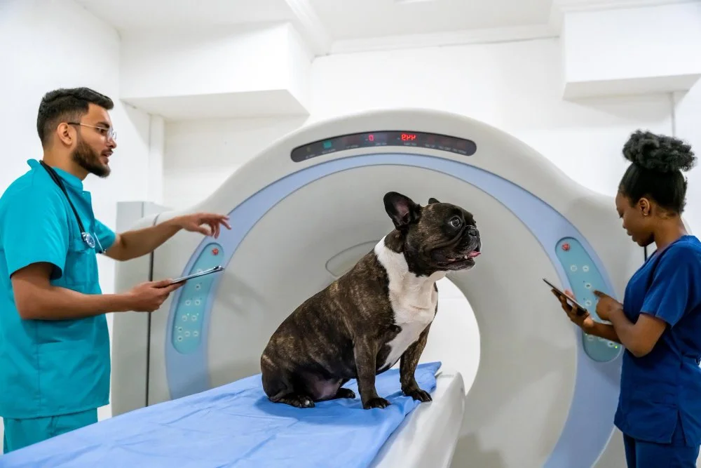
Magnetic Resonance Imaging (MRI) is one of the most advanced tools in veterinary medicine. Using a powerful magnet and radio waves, it creates highly detailed images of your pet’s internal organs, soft tissues, and nervous system—without the use of radiation.
A window into the nervous system
MRI is especially valuable for evaluating the brain and spine, offering the highest level of detail available in veterinary imaging. It allows doctors to visualize the brain’s structure, spinal cord, spinal nerves, and intervertebral discs, helping uncover causes of conditions such as seizures, weakness, pain, or changes in behavior.
Beyond the nervous system
MRI can also identify subtle or chronic musculoskeletal injuries that may not appear on X-rays or CT scans, including torn ligaments, muscle injuries, or joint inflammation. This makes MRI an important tool for diagnosing orthopedic issues and guiding long-term treatment.
Supporting long-term health decisions
In some cases, MRI findings can even help inform breeding programs or genetic counseling, especially when structural malformations are identified early.
By revealing such precise detail, MRI gives veterinarians the information they need to make accurate diagnoses and confident treatment plans—helping pets get relief sooner and avoid unnecessary procedures.