Radiographic Case Review: Left-Sided Congestive Heart Failure in an Elderly Chihuahua

An elderly Chihuahua was brought to SVI for a persistent cough that radiographs helped clarify.
Clinical Presentation:
An 8-year-old male neutered Chihuahua presented with a persistent wheezing cough of three months' duration. The dog exhibited a 4/6 left-sided systolic heart murmur, but no history of exercise intolerance or poor appetite was reported. During the radiographic examination, the patient became dyspneic, prompting concern for underlying cardiopulmonary disease.
Radiographic Assessment:
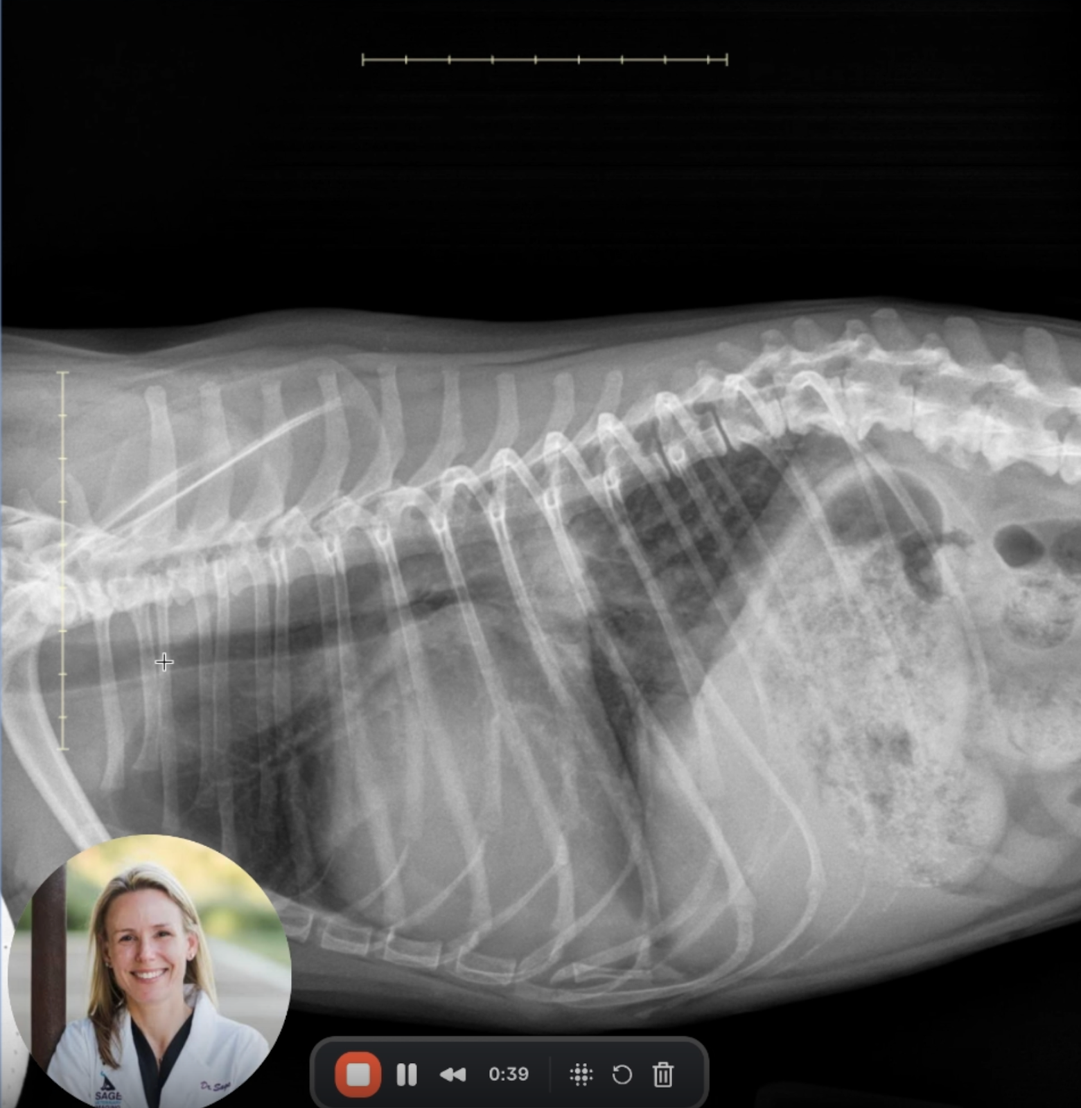
Image 1: Right Lateral View
Cardiac Silhouette: There is marked enlargement of the left atrium, evidenced by loss of the caudal cardiac waist and a soft tissue mass effect in the perihilar region.
Tracheal Deviation: Instead of the normal ventral deflection, the trachea runs parallel or slightly dorsal to the spine—a classic sign of left ventricular enlargement..
Pulmonary Parenchyma: There is a patchy increase in opacity within the caudodorsal lung fields, with a hypervascular appearance suggestive of early pulmonary venous congestion.
Additional Findings: Mineral debris is noted in the stomach (non-obstructive), the liver is at the upper limits of normal size, and chronic degenerative changes (disc space narrowing, endplate sclerosis, spondylosis) are seen in the caudal thoracic and cranial lumbar spine.
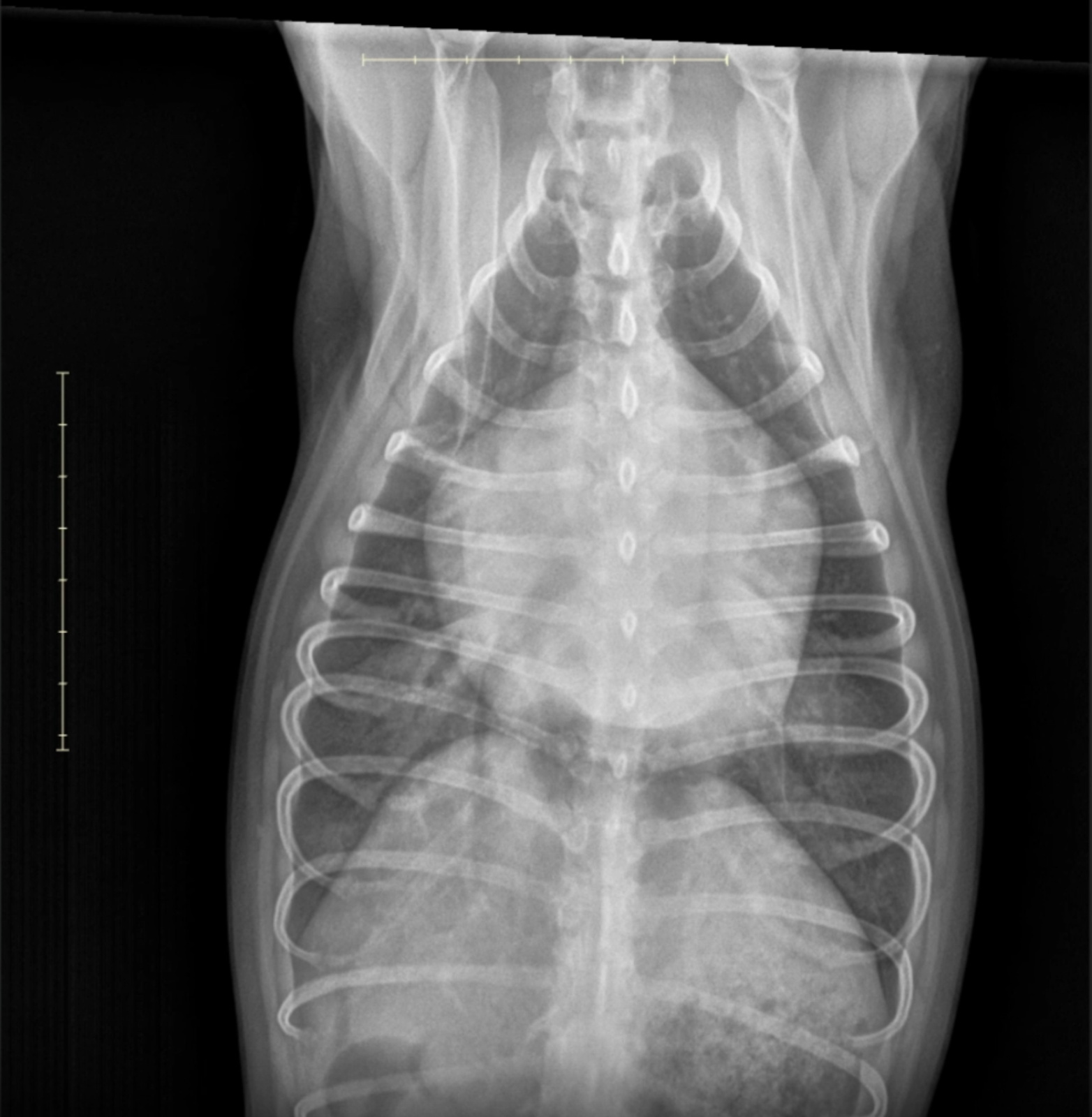
Image 2: Ventrodorsal (VD) View
Cardiac Findings: A clear "double opacity" sign and enlargement of the left atrial silhouette is visible, displacing adjacent bronchi and creating a “bow-legged cowboy” splaying of the mainstem bronchi.
Pulmonary Pattern: A distinct alveolar pattern is seen, most severe in the perihilar region, consistent with cardiogenic pulmonary edema.
Vasculature: Pulmonary veins appear mildly distended relative to arteries—supporting a post-capillary (left-sided) cause of congestion.
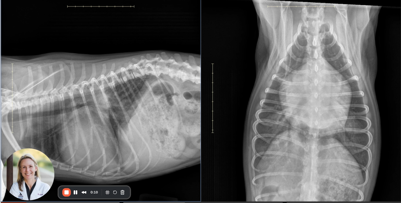
Image 3: Composite Overview
This comparison reaffirms the findings described above and offers a helpful orientation of the orthogonal projections. The right lateral and VD images together confirm both left atrial and ventricular enlargement, vascular congestion, and pulmonary edema.
Interpretation and Differentials:
The radiographic features support a diagnosis of:
Severe left-sided cardiac enlargement, including both the left atrium and left ventricle.
Pulmonary venous distension.
Perihilar alveolar and interstitial infiltrates, characteristic of cardiogenic pulmonary edema.
Together, these findings are consistent with left-sided congestive heart failure (CHF). The presumed underlying cause is chronic myxomatous mitral valve disease (MMVD) with secondary mitral regurgitation, common in small-breed, geriatric dogs.
Educational Notes for Clinical Practice:
Radiographic Hallmarks of Left-Sided CHF:
Elevation or straightening of the trachea (due to left ventricular enlargement).
Loss of the caudal cardiac waist and mainstem bronchial splaying (due to left atrial enlargement).
Pulmonary venous distension > artery size.
Perihilar interstitial or alveolar opacity—typically bilateral but more severe dorsally.
Differential Diagnosis Tips:
Non-cardiogenic pulmonary edema (e.g., electrocution, seizure, ARDS) usually has a caudodorsal distribution but lacks cardiomegaly.
Primary pulmonary disease typically lacks concurrent vascular and cardiac signs.
Tracheal elevation alone must be interpreted cautiously; position and obesity can mimic cardiomegaly.
Follow-Up Recommendations:
Thoracic point-of-care ultrasound to assess left atrial size, pulmonary B-lines.
Echocardiography for mitral valve anatomy and functional assessment.
NT-proBNP testing may support cardiogenic origin in equivocal cases.
Conclusion:
This case offers a textbook radiographic representation of left-sided congestive heart failure secondary to chronic mitral valve disease, a critical condition in geriatric small-breed dogs. Early recognition of the radiographic signs—particularly left atrial enlargement and pulmonary edema patterns—can greatly enhance diagnostic confidence in general practice.