
20 Reasons Your Vet Might Recommend a CT Scan After an Ultrasound
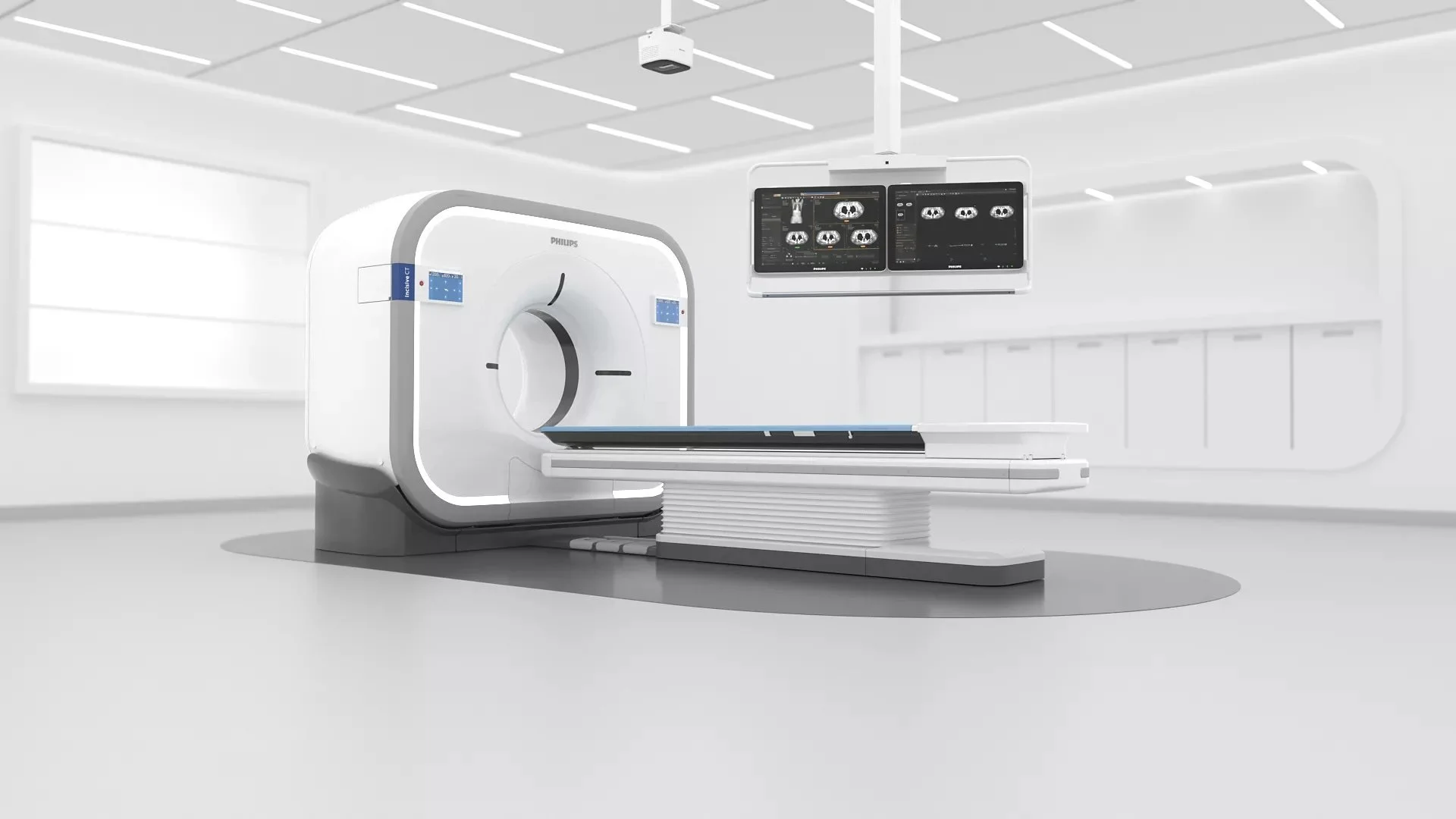
Our state-of-the-art CT scanner delivers fast, high-resolution 3D images, helping your vet see what ultrasound can’t and make confident, life-changing decisions for your pet.
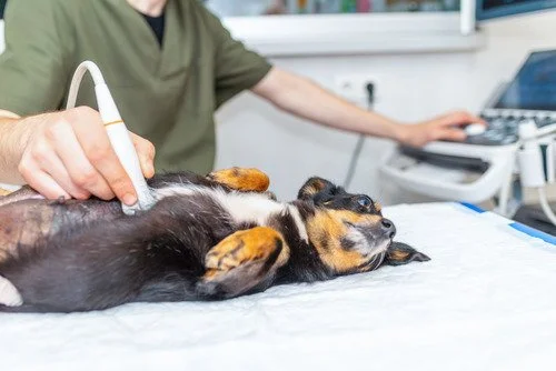
Ultrasound is one of the most common first steps in veterinary diagnostics. It’s quick, non-invasive, and great for capturing real-time images of your pet’s internal organs.
But sometimes, an ultrasound only gives part of the picture. When your vet needs more detail — or a clearer look at what’s really going on — a CT scan (computed tomography) may be the next best step.
At Sage Veterinary Imaging, we often see pets referred for a CT after an ultrasound has already been performed. That’s because while ultrasound is wonderful for soft tissue imaging, CT scans provide 3D detail, depth, and precision that ultrasound simply can’t match.
This article will help you understand why your vet might recommend a CT scan, what kinds of conditions require it, and how it helps ensure your pet gets the best possible care.
Why CT Is a Necessary Next Step
Think of ultrasound as a flashlight — it shows what’s immediately in front of it. CT, on the other hand, is like switching on a full 3D spotlight.
CT scans help your vet:
See inside or around a mass to understand if it’s cancerous or invasive
Detect tiny gas pockets, blood vessels, or bleeding sources that ultrasound may miss
Get surgical planning images for safer, more precise procedures
Map organs, bones, and blood flow in clear 3D detail
In many cases, CT gives the answers ultrasound simply can’t which can be the difference between guessing and knowing exactly what’s happening.
20 Conditions Where CT Should Always Follow Ultrasound
Ultrasound is often the first step in finding answers, but when results are unclear or incomplete, a CT scan can reveal the full picture.
If your pet’s ultrasound showed any of these findings, a CT scan may be the next logical step:
Splenic or Liver Masses
Ultrasound can find a mass — but CT helps determine if it’s spreading, connected to nearby vessels, or safe to remove surgically.Internal Bleeding (Hemoabdomen)
Ultrasound shows fluid; CT pinpoints where it’s coming from.Unclear or “Floating” Mass
If the ultrasound can’t tell where a mass starts or ends, CT reveals the exact origin and structure.Infections or Abscesses
CT helps locate gas pockets, infected areas, or abscesses, and guides your vet on the safest way to treat them.Chest or Lung Changes
When ultrasound shows fluid or thickening in the chest, CT helps distinguish between inflammation, fibrosis, or cancer.Dilated Bile Duct (CBD)
CT provides a full map to identify blockages — like stones, strictures, or tumors.Urinary or Urethral Concerns
Because ultrasound can’t easily see through bone, CT offers the best view of the pelvic canal and urethra.Cancer Staging
CT is the gold standard for checking spread to lungs, lymph nodes, or bones.Enlarged Lymph Nodes
CT shows whether lymph nodes are isolated or part of a wider condition.Lung or Mediastinal Mass
CT confirms what’s mass vs. fluid and maps exact size and spread.Suspected Liver Shunt
When ultrasound shows a small liver with high bile acids, CT angiography is the best way to locate shunts.Intestinal or Colonic Issues
CT differentiates between torsion, blockage, or inflammation.Diaphragmatic Hernia
CT reveals which organs have moved and how large the defect is.Dilated Veins (CVC or Hepatic)
When ultrasound shows abnormal vessels, CT uncovers possible causes like thrombosis or vascular shunts.Pancreatitis or Suspected Tumor
CT identifies inflammation, hidden masses, or blood clots in surrounding vessels.Pulmonary Nodules or Spots
CT evaluates their size, number, and if they’ve spread to lymph nodes.Unexplained Illness
When lab work and ultrasound don’t match your pet’s symptoms, CT often reveals hidden disease or infection.Pelvic Mass or Deep Swelling
CT provides clarity when ultrasound can’t see through the pelvis.Complex or Recurrent Infections
Especially when fluid keeps returning — CT helps find what’s been missed.Any Finding That Doesn’t Add Up
If your vet says, “something looks off, but I can’t tell exactly what,” CT is often the best next step.
Quick Reference: When to Refer from Ultrasound to CT
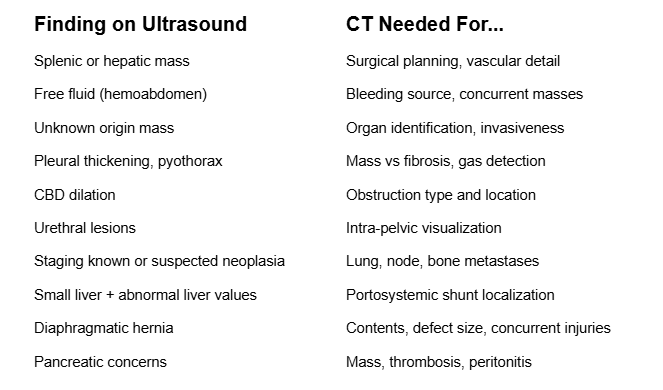
A quick reference guide showing when a CT scan should follow an ultrasound — helping your vet move from initial findings to a complete, confident diagnosis.
Why a CT Scan Can Be the Best Next Step
If your vet recommends a CT scan, it’s not about doing “extra testing” — it’s about getting answers that matter. Every minute counts when your pet’s health is at stake, and CT often provides the clarity needed to choose the right treatment path.
An ultrasound is an incredible tool, but it only shows part of the story. When your vet needs more detail, or when something serious is suspected, a CT scan can make the difference between a vague suspicion and a confident diagnosis with a clear plan of care.
How Sage Veterinary Imaging Helps You See the Full Picture

Every visit begins in a calm, pet-friendly space designed to help you and your companion feel at ease from the moment you walk in.
At Sage Veterinary Imaging, our priority is to help your care team see the whole picture — not just part of it. We use advanced CT technology designed specifically for pets, giving your veterinarian precise, 3D images that support faster, safer, and more confident decision-making.
If any of the 20 ultrasound findings we listed above apply to your pet, or if you’re unsure whether a CT scan is the right next step, ask your vet or reach out to our team. We’re always happy to review your pet’s case, explain what to expect, and help you make the most informed choice for your furry family member.