
Case Study: MRI Evaluation of Chronic Shoulder Pain in a Border Collie
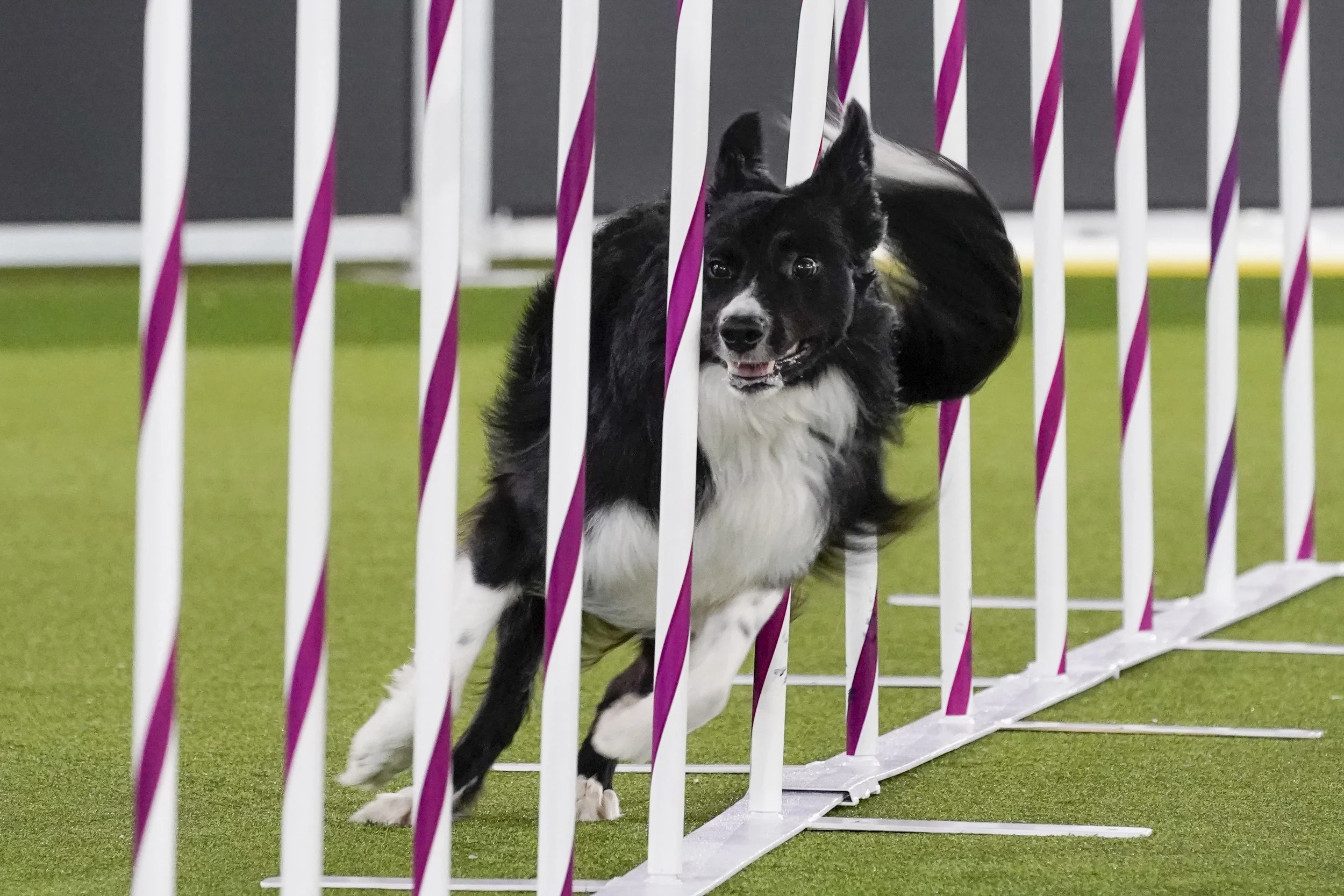
Meet Kal
Kal is a 4-year-old male intact Border Collie, an active and highly athletic dog participating in agility and outdoor activities. He presented with a history of intermittent right forelimb lameness, most noticeable after exercise.
Listen and watch as Dr. Sage walks through this complex case: View the Case Study
Clinical History
Kal’s lameness had been ongoing for over two months. Despite multimodal therapy, including chiropractic adjustments, laser therapy, and anti-inflammatory medications—his symptoms persisted. While there were periods of improvement, discomfort reliably returned following activity.
Radiographs revealed mild cervical spine changes and minimal carpal osteoarthritis. However, these findings did not adequately explain the degree or pattern of lameness, prompting further investigation.
Why MRI Was Recommended
Magnetic Resonance Imaging (MRI) was performed to evaluate both the cervical spine and shoulder joints.
MRI is particularly valuable in these cases because it allows for detailed assessment of soft tissues such as tendons, muscles, and joint structures, as well as the detection of inflammation and fluid. This level of detail is not achievable with radiographs alone and is critical in diagnosing subtle or complex musculoskeletal injuries.
Imaging Findings
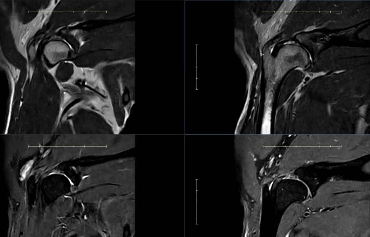
MRI scans of Kal's shoulders showing bilateral pathology. Findings include biceps tendinopathy with fluid in the bicipital tendon sheath (both sides), subscapularis tendon injury, and synovitis, with more advanced changes on the right.
Cervical Spine
Mild degenerative changes were noted at the C4–5 and C5–6 intervertebral discs, consistent with early disc dehydration. There was no evidence of spinal cord compression or neurologic abnormality.
These findings were considered incidental and unlikely to be the source of Kal’s lameness.
Left Shoulder
Findings in the left shoulder were mild but clinically relevant:
Subtle enlargement of the supraspinatus tendon at its insertion
Mild increased signal within the biceps tendon, indicating early inflammation
Small volume of fluid within the bicipital tendon sheath
These changes are consistent with low-grade tendinopathy and mild inflammation.
Right Shoulder (More Significant Findings)
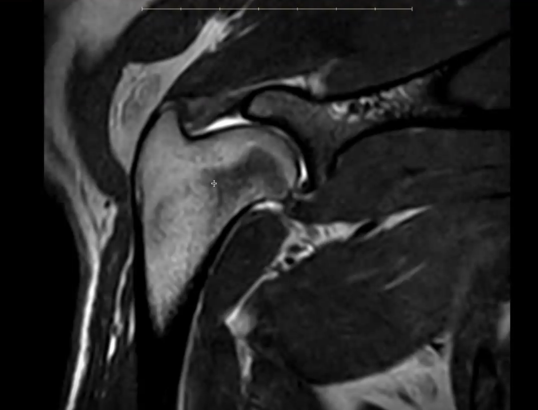
Close-up MRI view of Kal's shoulder joint, showing the structures involved in his tendon and joint inflammation.
More pronounced abnormalities were identified in the right shoulder:
Increased signal and structural changes within the subscapularis tendon, extending into the adjacent muscle
Contrast enhancement within the joint, consistent with synovitis (joint inflammation)
Intratendinous signal change in the biceps tendon
Moderate fluid accumulation within the bicipital tendon sheath
Together, these findings indicate more advanced tendon injury and active joint inflammation.
Interpretation
The MRI findings support bilateral shoulder pathology, with greater severity on the right side.
Kal’s lameness is best explained by a combination of:
Biceps tendinopathy (both shoulders)
Subscapularis tendon injury (right > left)
Synovitis (joint inflammation)
These structures play a key role in stabilizing the shoulder joint. When inflamed or injured, they can lead to pain, reduced performance, and intermittent lameness, particularly in highly active dogs.
Importantly, bilateral involvement can make lameness appear inconsistent, as dogs often shift weight between limbs to compensate.
Why This Matters
This case illustrates a common but important clinical reality: chronic lameness in athletic dogs is often multifactorial and driven by soft tissue injury rather than bone abnormalities.
Without MRI, these changes—particularly tendon pathology and joint inflammation—would likely have remained undetected.
Recommendations
Management should focus on addressing both tendon injury and joint inflammation. A multimodal approach is typically recommended, which may include:
Controlled rehabilitation and physical therapy
Activity modification to reduce repetitive strain
Anti-inflammatory or pain management strategies
Consideration of regenerative therapies (e.g., PRP or stem cell therapy)
Follow-up evaluation is important to monitor progress and guide return to activity.
Summary
MRI revealed bilateral shoulder tendon injury and joint inflammation, with more advanced disease affecting the right shoulder. These findings correlate closely with Kal’s history of activity-associated lameness and highlight the importance of advanced imaging in diagnosing complex orthopedic conditions.