
MRI or CT? A Practical Guide for Vets Navigating Neurologic and Orthopedic Cases
When Imaging Isn't Optional, But the Modality Is
Choosing the right scan depends on your dog’s symptoms and what your vet needs to see.
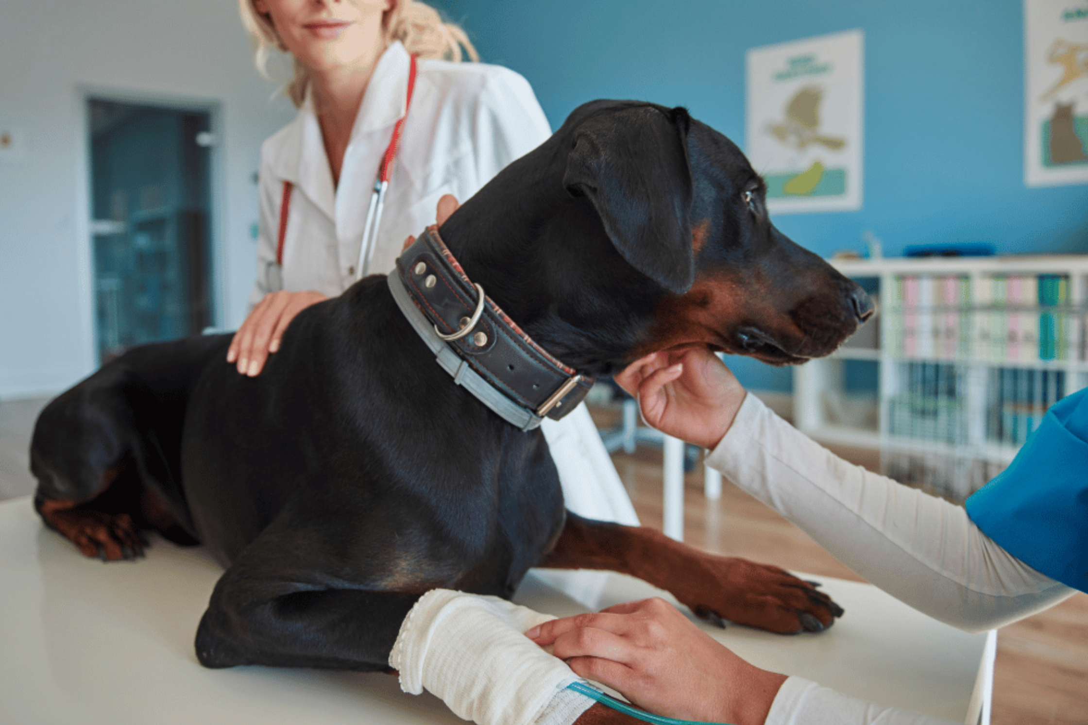
When a patient presents with unexplained lameness, spinal pain, seizures, or cranial nerve deficits, the critical question isn’t whether to image, but which modality to use: MRI or CT?
This decision directly impacts diagnostic accuracy, treatment speed, client trust, and total cost of care. At Sage Veterinary Imaging (SVI), we assist referring veterinarians in making this choice every day using both 3 Tesla MRI and 128-slice CT scanners on-site. This guide distills that expertise into actionable insights you can use in practice.
MRI vs. CT in Veterinary Medicine: Understanding Core Strengths
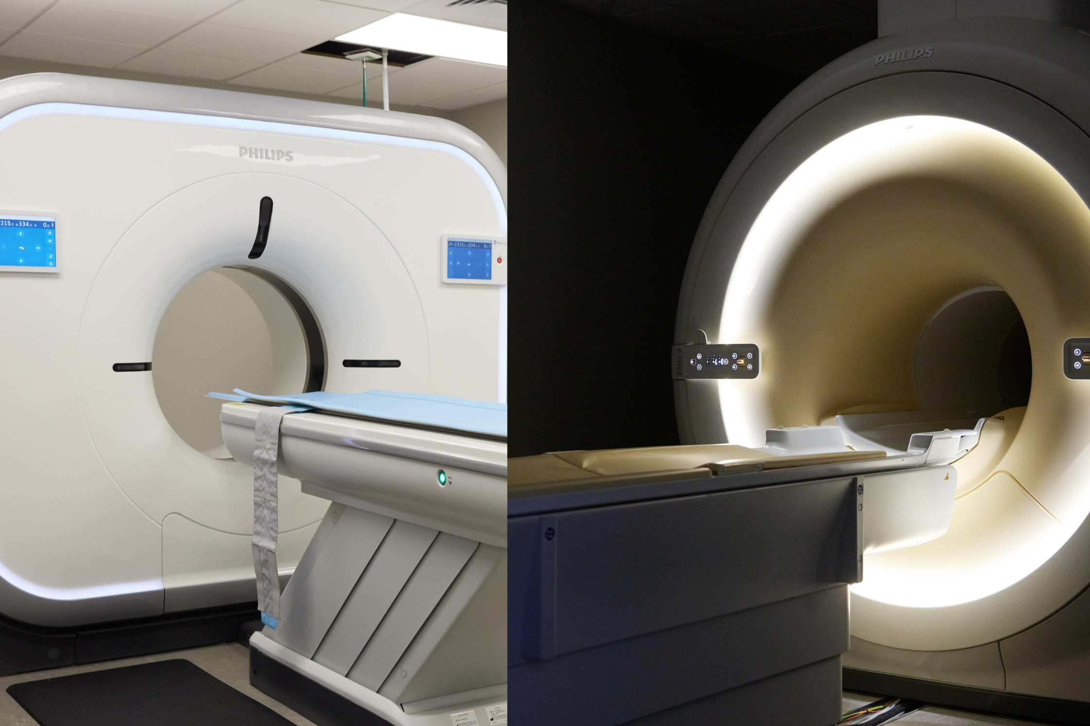
SVI uses high-resolution CT (left) and 3T MRI (right) machines to give pets the same advanced imaging used in human hospitals.
What MRI Does Best
Magnetic Resonance Imaging (MRI) uses a high-field magnet and radiofrequency pulses to produce detailed soft tissue images. It is unparalleled for:
Central nervous system (CNS) imaging – Detects spinal cord lesions, brain tumors, hydrocephalus.
Disc disease – Ideal for diagnosing Hansen Type I and II IVDD.
Seizure evaluations – Differentiates idiopathic epilepsy from structural causes.
Vestibular and cranial nerve dysfunction – Superior for brainstem, CN VII/VIII evaluation.
Soft tissue lameness – Shoulder instability, cruciate ligament injury, biceps tendinopathy.
Post-op neuro complications – Evaluates spinal cord integrity and surgical success.
Sage Note: Our 3T MRI offers resolution and contrast capabilities far beyond 1.5T systems, critical when assessing small lesions, cranial nerves, or subtle parenchymal abnormalities.
What CT Excels At
Computed Tomography (CT) uses X-ray technology to produce fast, high-resolution cross-sectional images, ideal for:
Bony lesions – Excellent for identifying osteolysis, periosteal reactions, fracture lines.
Dental and mandibular pathology – Tooth root abscesses, jaw tumors.
Nasal and sinus disease – Detects turbinate destruction, mineralized masses.
Thoracic and lung imaging – Pulmonary nodules, metastasis, pneumothorax
Ear disease – Visualizes tympanic bullae and middle ear fluid.
Trauma – Rapid screening for fractures, hemorrhage, or thoracoabdominal injury.
Oncology – Margin delineation for bone tumors, surgical planning.
Sage Note: Our 128-slice CT scanner completes full-body studies in under 30 seconds—ideal for unstable, anesthetic-sensitive patients.
Quick Reference Table: MRI vs. CT for Common Clinical Scenarios
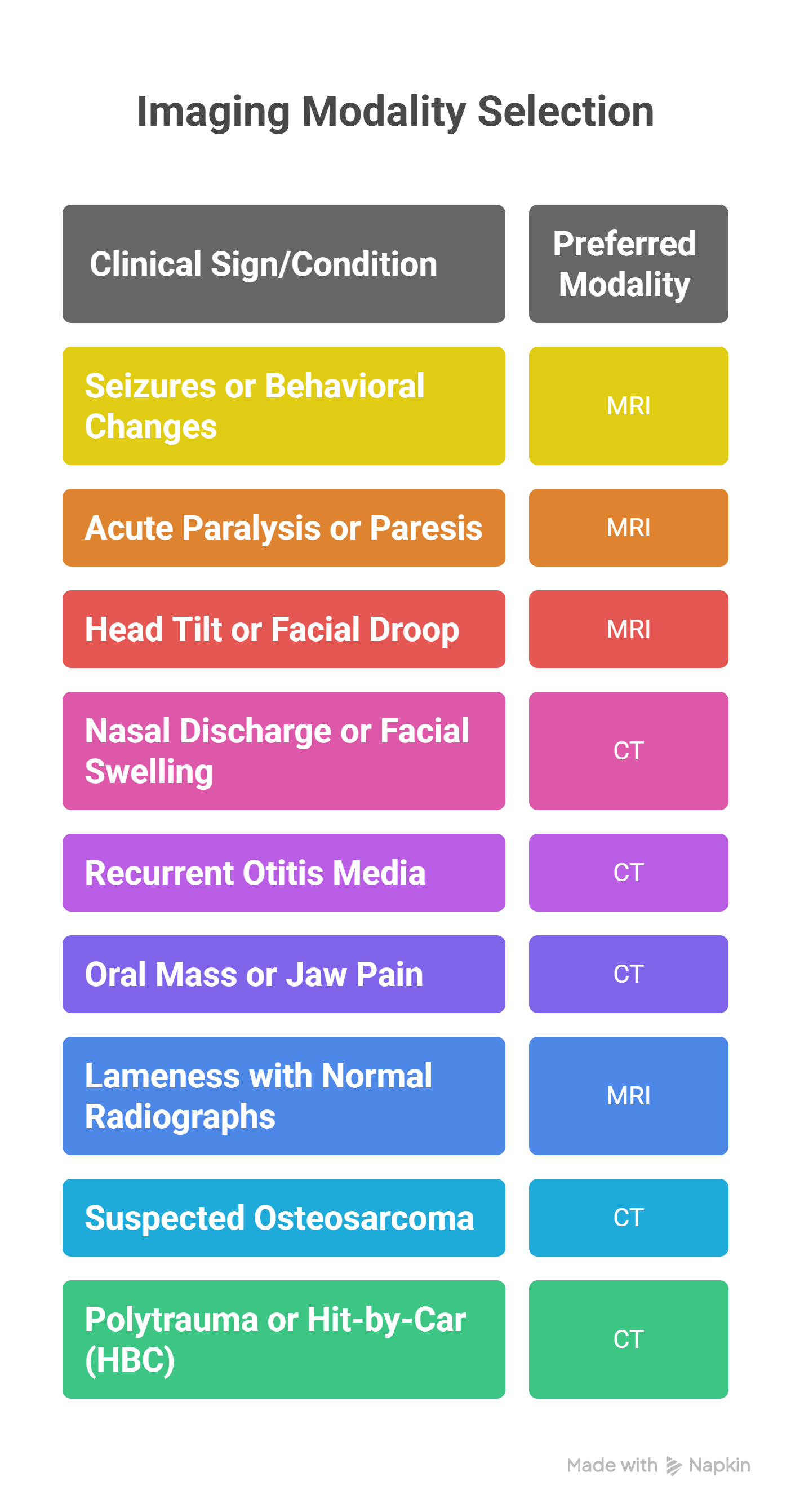
This quick guide helps clinicians match symptoms to the most informative modality.
Why Not Both? When MRI and CT Work Together
Some cases benefit from dual-modality imaging, especially when both soft tissue and bony components are involved:
Craniofacial trauma: CT identifies fractures; MRI assesses brain damage.
Limb mass staging: CT for bone margins, MRI for soft tissue extension.
Post-surgical neuro cases: MRI evaluates spinal cord; CT confirms implant alignment.
When medically safe, we coordinate CT and MRI under a single anesthesia event, reducing risk, cost, and case complexity.
Imaging Cost Considerations
Historically, MRI has been viewed as “too expensive” for many clients. That’s changing.
Pet insurance frequently covers MRI for neurologic indications.
At SVI, MRI and CT pricing is tiered by study type, not by hourly use.
For cost-sensitive cases, CT offers a rapid and affordable first-look, especially for bony pathologies or masses.
Collaboration Over Guesswork
We don’t just scan. We consult. SVI encourages referring veterinarians to reach out when imaging decisions aren’t clear-cut.
We can:
Review case history and suggest ideal imaging sequences
Recommend sedation vs general anesthesia
Discuss staging plans for oncology or trauma cases
Book same-day or next-day appointments when needed
Final Takeaway: Imaging That Supports, Not Replaces, Your Diagnosis
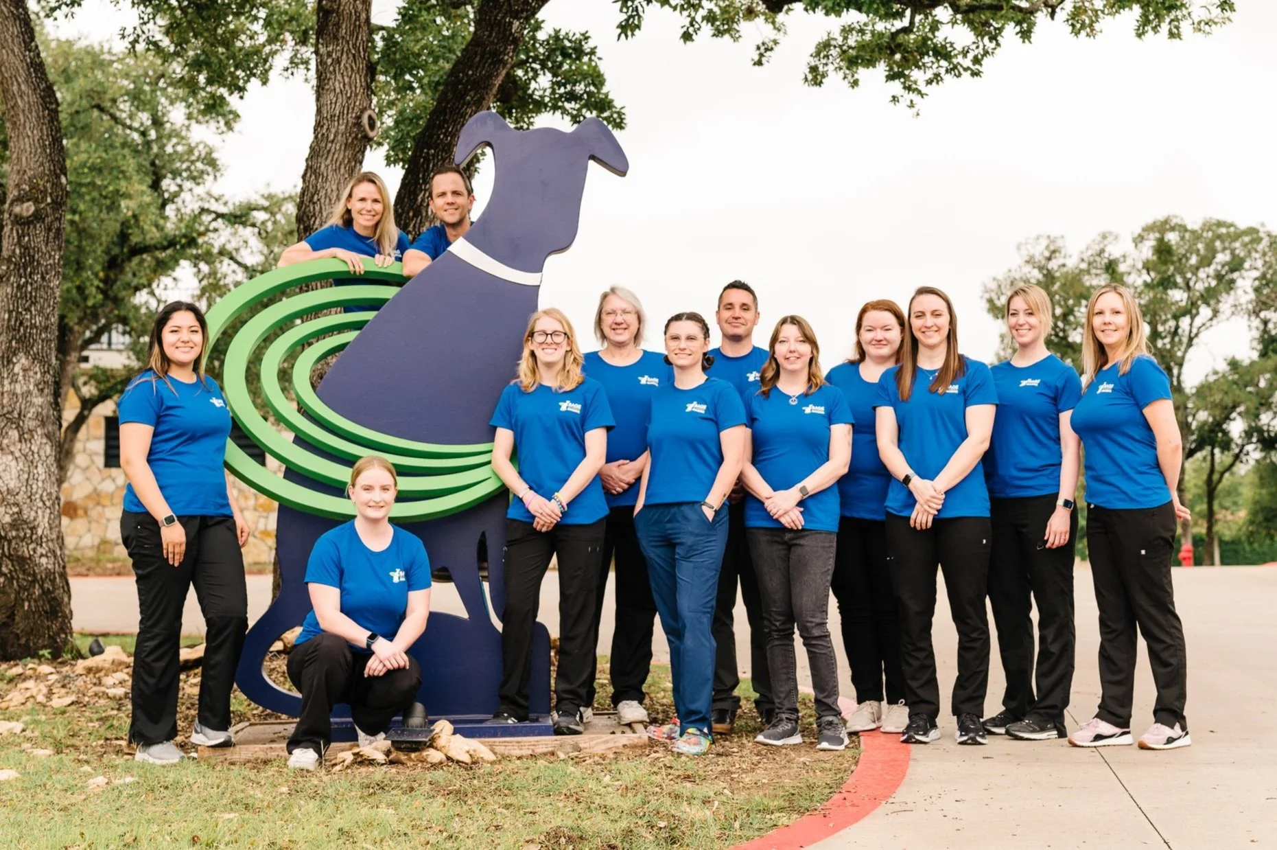
Meet the compassionate team behind Sage Veterinary Imaging, dedicated to delivering fast, advanced diagnostics for your pet.
Advanced imaging is a clinical extension, not a replacement, for your judgment. CT and MRI should empower you to:
Confirm or rule out differentials
Plan surgery or treatment with precision
Educate clients with confidence and clarity
At Sage Veterinary Imaging, we’re here to make that easier.
Need Input on a Case?
Contact us for a consult. We’ll help you choose the right imaging path, quickly, collaboratively, and with clinical integrity.