
Advanced CT and MRI imaging help veterinarians detect and evaluate serious conditions like hemangiosarcoma, providing critical information for diagnosis and treatment planning.
Hemangiosarcoma is an aggressive and often silent cancer in dogs, making early detection critically important. Unfortunately, many dogs show few signs until the disease is advanced.
For pet owners in Austin, Round Rock, and Central Texas, advanced imaging—particularly CT and MRI—plays a key role in identifying hemangiosarcoma earlier and helping veterinarians make informed treatment decisions. At Sage Veterinary Imaging in Round Rock, veterinarians refer patients for high-quality imaging to evaluate suspected cancers like hemangiosarcoma.
What Is Hemangiosarcoma?
Hemangiosarcoma is a malignant cancer of blood vessel cells. It most commonly affects:
The spleen
The heart (right atrium)
The liver
Skin or subcutaneous tissues
Because these tumors involve blood vessels, they are prone to rupture and internal bleeding, which can become life-threatening very quickly.
Why Early Detection Is Challenging
Hemangiosarcoma is often called a “silent disease” because:
Tumors can grow without obvious symptoms
Early signs are vague or mild
Internal tumors are not visible externally
Many dogs are diagnosed only after a sudden collapse or internal bleeding event, which limits treatment options.
Dogs at Higher Risk
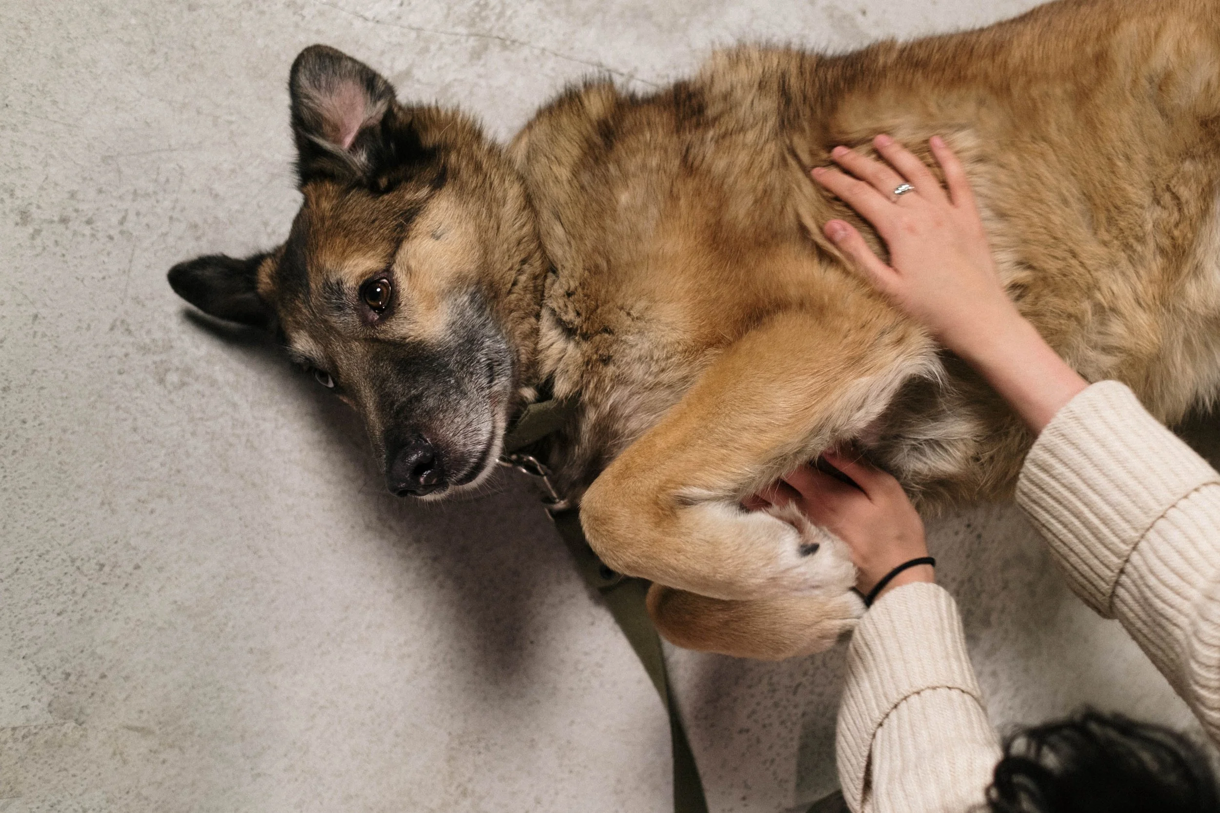
Many dogs with hemangiosarcoma show only subtle symptoms early in the disease process, making thorough veterinary evaluation an important first step.
While any dog can develop hemangiosarcoma, it is more common in:
Golden Retrievers
German Shepherds
Labrador Retrievers
Boxers
It typically affects middle-aged to older dogs.
Subtle Signs to Watch For
Early symptoms can be easy to miss but may include:
Lethargy or decreased energy
Reduced appetite
Intermittent weakness
Pale gums
Mild abdominal distension
In more advanced cases:
Sudden collapse
Difficulty breathing
Signs of internal bleeding
If your veterinarian suspects a serious underlying issue, advanced imaging may be recommended.
How CT and MRI Help Detect Hemangiosarcoma
Advanced imaging provides critical information that cannot be obtained through physical exams or basic X-rays.
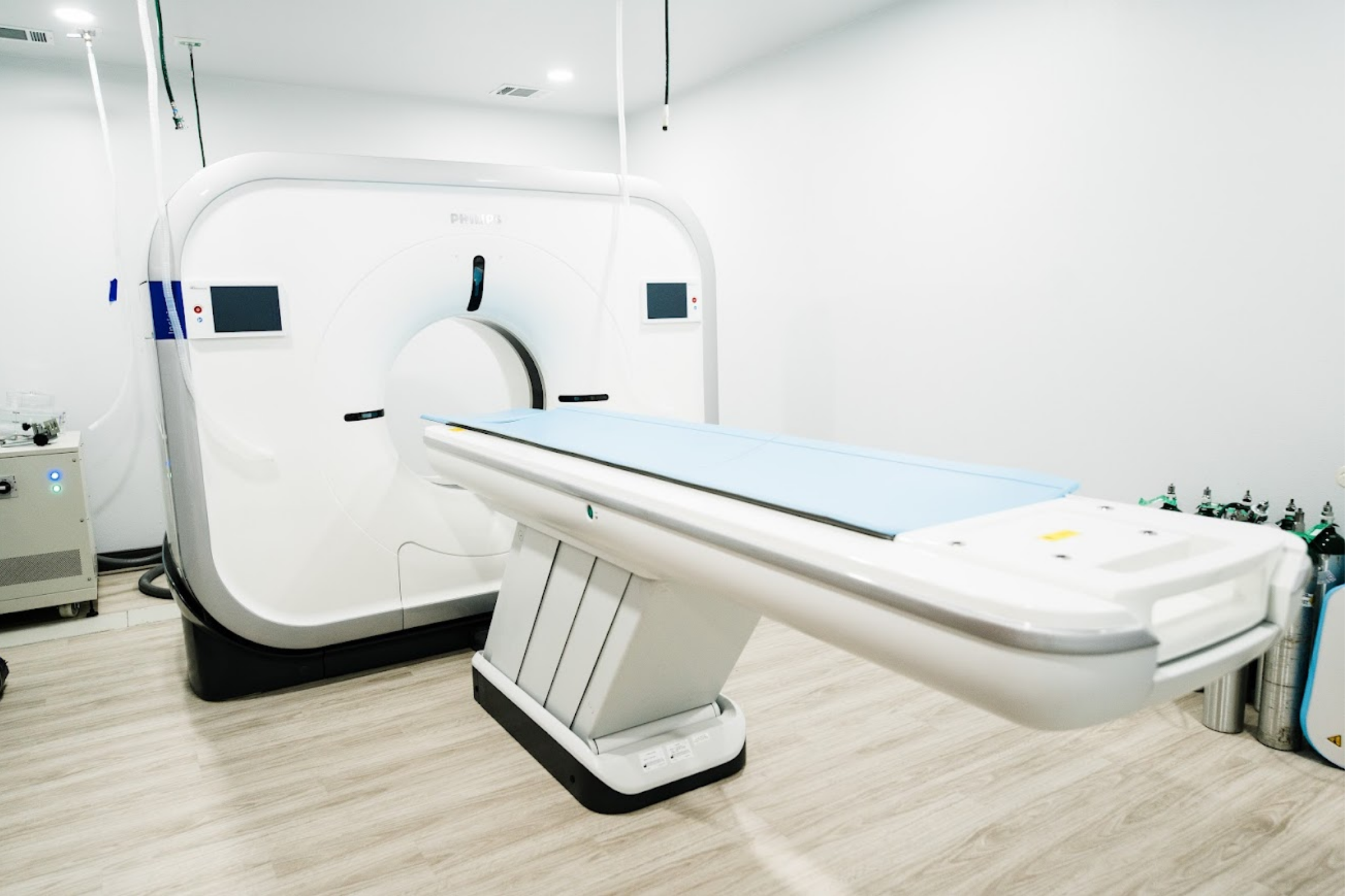
128-slice CT technology helps veterinarians detect tumors, evaluate cancer spread, and guide treatment planning for conditions such as hemangiosarcoma.
CT Scans for Cancer Detection and Staging
CT is often the first-line advanced imaging tool for suspected cancer because it is fast and highly detailed.
CT can:
Detect tumors in the spleen, liver, and lungs
Identify internal bleeding
Evaluate whether cancer has spread (metastasis)
Help guide surgical or treatment planning
At Sage Veterinary Imaging, 128-slice CT technology allows for rapid, high-resolution scans.
MRI for Soft Tissue and Complex Cases
MRI may be used in certain cases to:
Evaluate soft tissue involvement in greater detail
Assess neurological signs if cancer has spread
Provide additional clarity when CT findings are inconclusive
With 3T MRI technology, subtle abnormalities can be detected with greater precision.
Where to Get Advanced Imaging in Austin
Veterinary clinics throughout Austin, Round Rock, Cedar Park, Georgetown, Pflugerville, Leander, Kyle, and Buda often refer patients to Sage Veterinary Imaging in Round Rock for advanced cancer imaging.
Conveniently located along the Interstate 35 corridor, the facility provides accessible diagnostics for pets across Central Texas.
What to Expect During Imaging
Advanced imaging is carefully coordinated to ensure safety and accuracy.
A typical visit includes:
Veterinary coordination
Your veterinarian works with the imaging team to determine the most appropriate study.
Pre-procedure evaluation
Your dog is assessed to ensure they are safe for anesthesia if needed.
Imaging procedure
CT or MRI is performed using advanced equipment.
Radiologist interpretation
A board-certified veterinary radiologist reviews the images and provides a detailed report.
Your veterinarian will then discuss findings and treatment options with you.
Why Early Detection Matters
While hemangiosarcoma is aggressive, early detection can:
Identify tumors before rupture
Allow for more treatment options
Improve quality of life
Support better-informed decision-making
Even when a cure isn’t possible, earlier diagnosis can help guide timely and compassionate care.
A Collaborative Approach to Cancer Care
Sage Veterinary Imaging works closely with veterinarians across Central Texas to support early detection and staging of cancer.
The imaging team provides:
Advanced CT and MRI diagnostics
Expert radiologist interpretation
Clear, timely communication with your veterinarian
Your primary veterinarian remains responsible for treatment planning and ongoing care.
Helping Austin-Area Dogs Get Answers Sooner

Sage Veterinary Imaging provides advanced CT and MRI services for referring veterinarians throughout Austin and Central Texas.
Hemangiosarcoma can be difficult to detect—but advanced imaging gives veterinarians powerful tools to find and evaluate this disease earlier.
At Sage Veterinary Imaging in Round Rock, CT and MRI technology help provide clarity when it matters most.
If your veterinarian suspects a serious condition or recommends further evaluation, advanced imaging is available nearby to help provide answers and guide the next steps in your dog’s care.